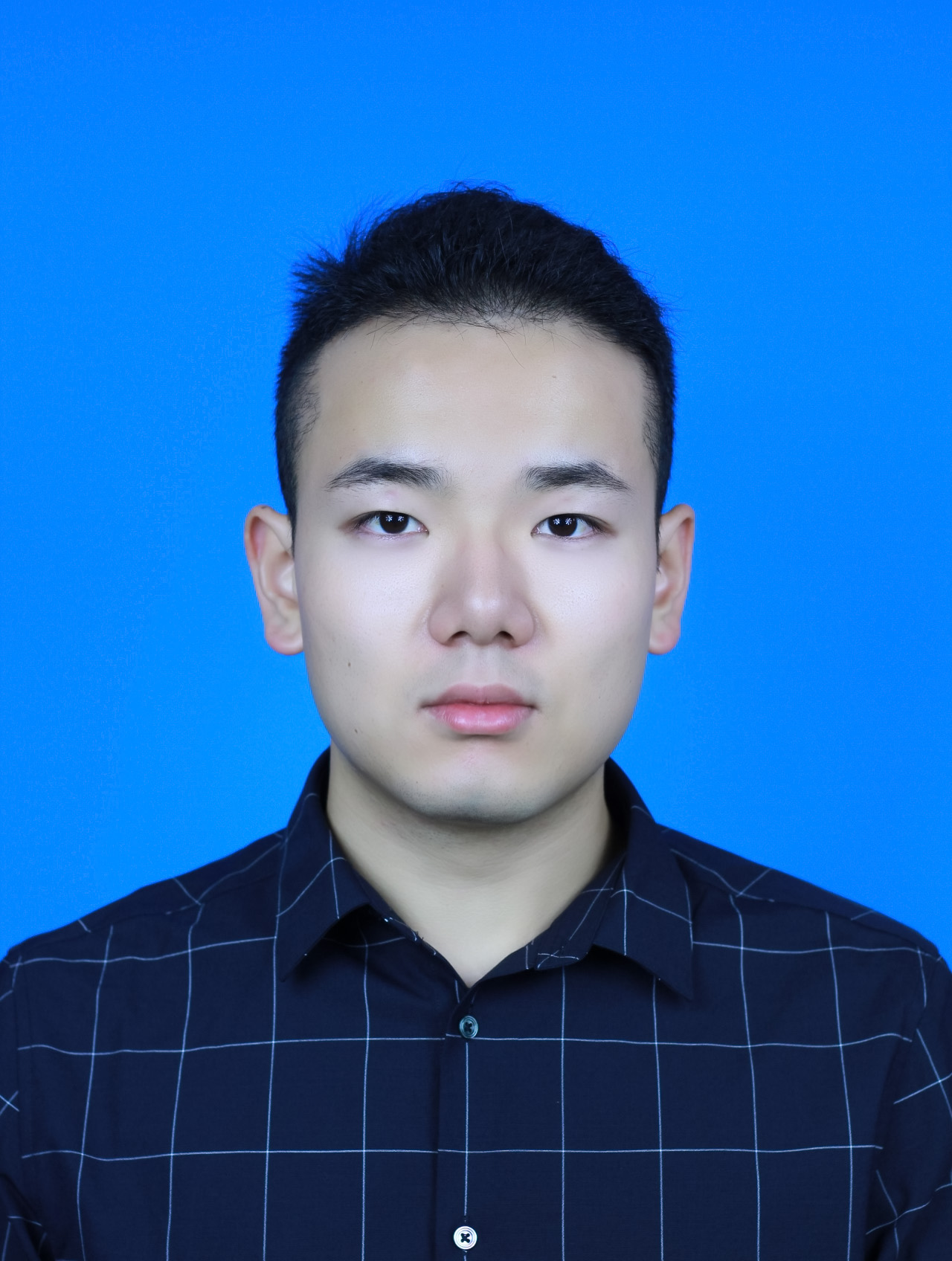
Wenhui Lei

Shanghai Jiaotong University (SJTU), Shanghai, China
lyc745307452@gmail.com
Research Interests
- Multi-Modality Learning;
- Foundation Models;
- Image Generation;
- Self-supervised Learning;
- Label Efficient Learning
News
- 2024.. One paper for Localization Foundation Model is accepted by MIA!
- 2023.. One paper for One-shot Segmentation is accepted by TMI!
- 2023.. One paper for Generalizable Medical Image Segmentation is accepted by MIA!
- 2023.. One paper for Effecient Subclass Learning is early accepted by MICCAI 2023!
- 2023.. One paper for Brain Organ-at-Risk Segmentation is accepted by Medical Physics!
- 2022.. One paper for Domain Adaptation is accepted by TMI!
- 2022.. One paper for Brain Structure segmentation is accepted by JBHI!
- 2022.. One paper for Head-and-Neck Organ-at-Risk Segmentation is accepted by Neurocomputing!
- 2022.. One paper for Gross Target Volume of Nasopharynx Cancer Segmentation is accepted by Neurocomputing!
- Won the MICCAI2021 Student Travel Award!
- 2021.. One paper for One-shot Localization is accepted by MICCAI 2021!
- 2019.. One paper for Interactive Segmentation is accepted by MICCAI LABELS 2019 as oral presentation!
Publications
- One-shot Weakly-Supervised Segmentation in Medical Images, IEEE Transactions on Medical Imaging, 2022. Lei Wenhui, Su Qi, et al.
- Contrastive Learning of Relative Position Regression for One-Shot Object Localization in 3D Medical Images, MICCAI (Early accepted, student travel award), 2021. Lei Wenhui, Wei, X., et al.
- Domain Composition and Attention for Unseen-Domain Generalizable Medical Image Segmentation, Accepted by MICCAI, 2021. Gu Ran, Zhang Jingyang, Huang Rui, Lei Wenhui, et al.
- Automatic Segmentation of Organs-at-Risk from Head-and-Neck CT using Separable Convolutional Neural Network with Hard-Region-Weighted Loss, Neurocomputing, 2021. Lei, W., Mei, H., et al.
- “DeepIGeoS-V2: Deep Interactive Segmentation of Multiple Organs from Head and Neck Images with Lightweight CNNs.” MICCAI Workshop LABELs(oral), 2019. Lei, W., Wang, H., et al.
- Automatic Segmentation of Gross Target Volume of Nasopharynx Cancer using Ensemble of Multiscale Deep Neural Networks with Spatial Attention, Neurocomputing, 2021. Mei, H., Lei, W., et al.
- “A Weighted Ensemble Coarse-to-fine Framework for Myocardial Edema and Scars Segmentation” MICCAI Workshop MyoPS, 2020. Zhai, S., Gu, R., Lei, W., and Wang, G.,
- “CSAF-CNN: Cross-Layer Spatial Attention Map Fusion Network for Organ-at-Risk Segmentation in Head and Neck CT Images.” ISBI, 2020. Liu, Z., Wang, H., Lei, W., Wang, G.
- “High-and Low-Level Feature Enhancement for Medical Image Segmentation.” MICCAI Workshop MLMI, 2019. Wang, H, Wang, G., Xu, Z., Lei, W., Zhang, S.
Challenges
- ABCs 2020 challenge:
- Won the 2nd place in MICCAI 2020 Anatomical Brain Barriers to Cancer Spread (ABCs) Challenge task 1: segmenting 5 brain structures to use for automated definition of the clinical target volume (CTV) for radiotherapy treatment.
- Won the 2nd place in MICCAI 2020 ABCs task 2: segmenting 10 structures to use in radiotherapy treatment plan optimization.
- MyoPS 2020 challenge:
- Won the 1st place in MICCAI 2020 Myocardial Pathology Segmentation Combining Multi-sequence CMR Challenge: Proposed a weighted ensemble coarse-to-fine framework for myocardial edema and scars segmentation. It consists of:
- Coarse-to-fine model which first predict the cardiac structure area and then output a detailed prediction.
- A weighted ensemble model to integrate prediction from 2D network and 2.5D network.
- Won the 1st place in MICCAI 2020 Myocardial Pathology Segmentation Combining Multi-sequence CMR Challenge: Proposed a weighted ensemble coarse-to-fine framework for myocardial edema and scars segmentation. It consists of:
- StructSeg 2019 challenge:
- Won the 3rd place in MICCAI 2019 Automatic Structure Segmentation for Radiotherapy Planning (StructSeg) Challenge task 1 (head and neck organ). The method is mainly composed of:
- Segmental Linear Functions (SLFs) transform the intensity of CT images to make multiple organs more distinguishable than existing methods.
- A novel 2.5D network specially designed for dealing with clinic H&N CT scans with anisotropic spacing.
- Our proposed attention to hard voxels (ATH) uses a voxel-level hardness strategy, which is more suitable to dealing with some hard regions despite that its corresponding class may be easy. The paper describing above methods is under revision of Neurocomputing (IF=4.438).
- Won the 2nd place in StructSeg task2 (head and neck tumor): The method is mainly composed by:
- Cropping data in large and small image mode for ensemble of features in different scales.
- A variant of the U-Net, in which each block was preceded by a ‘Project Excite’ (PE) block. It can recalibrate feature maps spatial-wisely on 3D volumetric images. The paper describing above methods is accepted by Neurocomputing.
- Won the 3rd place in MICCAI 2019 Automatic Structure Segmentation for Radiotherapy Planning (StructSeg) Challenge task 1 (head and neck organ). The method is mainly composed of:
Education
- Shanghai Jiao Tong University (SJTU), Shanghai, China
- PhD of Electronic Information, 2021.09 - Now
- University of Electronic Science and Technology of China (UESTC), Sichuan, China
- Master of Computer Vision and Image Processing, 2018.09 – 2021.07
- University of Electronic Science and Technology of China (UESTC), Sichuan, China
- Bachelor of Industrial Engineering, 2014.09 – 2018.09